Lower Back Organ Anatomy Diagram - Human midsection with internal organs Stock Photo - Alamy
Lower Back Organ Anatomy Diagram - Human midsection with internal organs Stock Photo - Alamy. The back supports the weight of the body, allowing for flexible movement while protecting vital organs and nerve structures. Lower back organ anatomy diagram / human back anatomy female back muscle anatomy human back diagram organs massage. This diagram depicts lower back anatomy diagram 744×1125.human anatomy diagrams show internal organs, cells, systems, conditions, symptoms and sickness information and/or tips for healthy living. The lumbar region of the spine, more commonly known as the lower back, is situated between the thoracic, or chest, region of the spine, and the sacrum. As you can see, there are also have a spine of scapula deltoid, triceps brachii, latissimus dorsi.
ads/bitcoin1.txt
7 photos of the human body organs in lower back. This article looks at the anatomy of the back, including bones, muscles. It is particularly interesting for physiotherapists. This diagram depicts lower back anatomy diagram 744×1125.human anatomy diagrams show internal organs, cells, systems, conditions, symptoms and sickness information and/or tips for healthy living. Diagram of a female lower back 4 photos of the diagram of a female lower back diagram of lower back and hips, diagram of lower back muscles and ligaments, diagram of lower back organs, lower back anatomy diagram, lower back diagram bones, lower back diagram pelvis, human anatomy, diagram of lower back and hips, diagram …
Pin on An anatomy lab from i.pinimg.com It is the strongest and most prominent part of the lower extremity, thus a personal favourite for fitness enthusiasts to showcase. The lumbar spine is the lower back that begins below the last thoracic vertebra (t12) and ends at the top of the sacral spine, or sacrum (s1). The abdomen contains all the digestive organs, including the stomach, small and large intestines, pancreas, liver, and gallbladder. Related posts of human anatomy female lower back muscle anatomy triceps. Each lumbar spinal level is numbered from top to bottom—l1 through l5, or l6. Lower back organ anatomy diagram / human back anatomy female back muscle anatomy human back diagram organs massage. The major organs of the abdomen include. It has an upper extremity, a shaft, and a lower.
The scaffold of the thigh is provided by the femur, the only bone of this region and the longest bone in the body.
ads/bitcoin2.txt
Related posts of female body back side anatomy skeleton bones diagram. Each bone is a complex living organ that is made up of many cells, protein fibers, and minerals. Each lumbar spinal level is numbered from top to bottom—l1 through l5, or l6. The back is the body region between the neck and the gluteal regions. As you can see, there are also have a spine of scapula deltoid, triceps brachii, latissimus dorsi. Diagram of a female lower back 4 photos of the diagram of a female lower back diagram of lower back and hips, diagram of lower back muscles and ligaments, diagram of lower back organs, lower back anatomy diagram, lower back diagram bones, lower back diagram pelvis, human anatomy, diagram of lower back and hips, diagram … They help to bend the back to one side or the other. It comprises the vertebral column (spine) and two compartments of back muscles; It is particularly interesting for physiotherapists. It is the strongest and most prominent part of the lower extremity, thus a personal favourite for fitness enthusiasts to showcase. Doctors usually list dozens of organs, though the definition of an organ varies from expert to expert. Bones of the pelvis and lower back. The major organs of the abdomen include.
The lumbar and sacrum region make up the bone of the lower back anatomy. Muscles of the abdomen lower back and pelvis. The back supports the weight of the body, allowing for flexible movement while protecting vital organs and nerve structures. See more ideas about muscle anatomy, anatomy, body anatomy. This article looks at the anatomy of the back, including bones, muscles.
Kidney Pain - (Location, anatomy), lower back, Vs Back ... from healthool.com Lower back organ anatomy diagram / human back anatomy female back muscle anatomy human back diagram organs massage. Most organs play a role in organ systems, which work together to perform specific functions. It is the surface of the body opposite from the chest and the abdomen.the vertebral column runs the length of the back and creates a central area of recession. The quadratus lumborum muscles (orange, in the image above) are found in the lower back (also called the lumbar area). Anatomy organs body anatomy anatomy art anatomy and physiology human anatomy human body organs human body art human organ diagram human body science. The muscles that move the upper legs (thigh) there are many muscles that move the large bone of the thigh. The major muscles of the abdomen include the rectus abdominis in front, the external obliques at the sides, and the latissimus dorsi muscles in the back. The back supports the weight of the body, allowing for flexible movement while protecting vital organs and nerve structures.
Female reproductive organs of the lower torso.
ads/bitcoin2.txt
The lumbar and sacrum region make up the bone of the lower back anatomy. Posted on february 14, 2015 by admin. In women, various reproductive organs located in the pelvis may lead to lower right back pain. The back supports the weight of the body, allowing for flexible movement while protecting vital organs and nerve structures. The pain may make it hard to move or stand up straight. These sections are cervical (neck), thoracic (upper and middle back), lumbar (lower back), and sacrum (tailbone). Anatomy organs body anatomy anatomy art anatomy and physiology human anatomy human body organs human body art human organ diagram human body science. Collectively this region is called the vulva. Immune and lymphatic systems of the lower torso. Bones of the pelvis and lower back. The back is the body region between the neck and the gluteal regions. As you can see, there are also have a spine of scapula deltoid, triceps brachii, latissimus dorsi. Each bone is a complex living organ that is made up of many cells, protein fibers, and minerals.
Diagram of a female lower back 4 photos of the diagram of a female lower back diagram of lower back and hips, diagram of lower back muscles and ligaments, diagram of lower back organs, lower back anatomy diagram, lower back diagram bones, lower back diagram pelvis, human anatomy, diagram of lower back and hips, diagram … These organs are held together loosely by connecting tissues. The quadratus lumborum muscles (orange, in the image above) are found in the lower back (also called the lumbar area). The muscles that move the upper legs (thigh) there are many muscles that move the large bone of the thigh. The lumbar spine is the lower back that begins below the last thoracic vertebra (t12) and ends at the top of the sacral spine, or sacrum (s1).
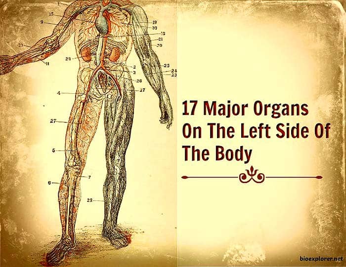
Human Body Diagram Left Side - Human Anatomy from www.bioexplorer.net For example, endometriosis is a common condition that may create sporadic, sharp pain in the pelvic area that may radiate to the lower right back. It is particularly interesting for physiotherapists. The human spine is composed of 4 sections of vertebrae. This human anatomy module is composed of diagrams, illustrations and 3d views of the back, cervical, thoracic and lumbar spinal areas as well as the various vertebrae. The muscles that move the upper legs (thigh) there are many muscles that move the large bone of the thigh. The major organs of the abdomen include. In this image, you will find an occipital bone, sternocleidomastoid, trapezius, deltoid in muscles of the lower back diagram. The breadth of the back is created by the shoulders at the top and the pelvis at the bottom.
The breadth of the back is created by the shoulders at the top and the pelvis at the bottom.
ads/bitcoin2.txt
Symptoms of low back pain. Each bone is a complex living organ that is made up of many cells, protein fibers, and minerals. The major organs of the abdomen include. It is particularly interesting for physiotherapists. The muscles that move the upper legs (thigh) there are many muscles that move the large bone of the thigh. Coming to the point of discussion, specific organs are present on the left and right side of the body, while some are located in the center, sharing both the orientations. Posted on february 14, 2015 by admin. The bones of the pelvis and lower back work together to support the body's weight, anchor the abdominal and hip muscles, and protect the delicate vital organs of the vertebral and abdominopelvic cavities. The skeleton acts as a scaffold by providing support and protection for the soft tissues that make up the rest of the body. Understanding lower back anatomy is key to understanding the root of lower back and hip pain. This human anatomy module is composed of diagrams, illustrations and 3d views of the back, cervical, thoracic and lumbar spinal areas as well as the various vertebrae. Collectively this region is called the vulva. The abdomen contains all the digestive organs, including the stomach, small and large intestines, pancreas, liver, and gallbladder.
ads/bitcoin3.txt
ads/bitcoin4.txt
ads/bitcoin5.txt
0 Response to "Lower Back Organ Anatomy Diagram - Human midsection with internal organs Stock Photo - Alamy"


0 Response to "Lower Back Organ Anatomy Diagram - Human midsection with internal organs Stock Photo - Alamy"
Posting Komentar